MBL

Anti-Fas (CD95) mAb

Product Code:
 
MBL-SY-001
Product Group:
 
Primary Antibodies
Supplier:
 
MBL
Host Type:
 
Mouse
Antibody Isotype:
 
IgM
Antibody Clonality:
 
Monoclonal
Antibody Clone:
 
CH-11
Regulatory Status:
 
RUO
Target Species:
 
Human
Applications:
  • Flow Cytometry
  • Western Blot (WB)
Shipping:
 
4°C
Storage:
 
-20°C
1 / 2
2 / 2

No additional charges, what you see is what you pay! *

CodeSizePrice
MBL-SY-001100 ul£366.00
Quantity:
Prices exclude any Taxes / VAT
Stay in control of your spending. These prices have no additional charges, not even shipping!
* Rare exceptions are clearly labelled (only 0.14% of items!).
Multibuy discounts available! Contact us to find what you can save.
This product comes from: United States.
Typical lead time: 10-14 working days.
Contact us for more accurate information.
  • Further Information
  • Documents
  • References
  • Show All

Further Information

Applications:
WB - 20 ug/mLm (chemiluminescence detection system) FCM - 20 ug/mL (final conc.) Other - 100 ng/mL????37?2???????????????, 15????????????????????
Background:
It is now widely accepted that apoptosis plays an important role in the selection of immature thymocytes and Ag-primed peripheral T cells. Fas antigen is a cell-surface protein that mediates apoptosis. It is expressed in various tissues including the thymus and has structural homology with a number of cell-surface receptors, including tumor necrosis factor receptor and nerve growth factor receptor.
Formulation:
0.5 mg/mL in PBS/50% glycerol, pH 7.2
Gene IDs:
Human: 355 Mouse: 14102
Immunogen Translated:
FS-7 Human diploid fibroblasts
Reactivity:
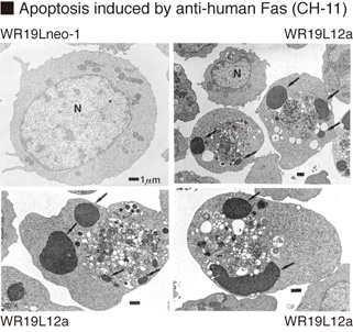
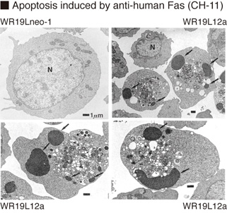
This antibody induces apoptosis in human cell lines expressing Fas antigen
Source:
This antibody was purified from ascites fluid using affinity chromatography. This hybridoma (clone CH-11) was established by fusion of mouse myeloma cell NS-1 with Balb/c mouse splenocyte immunized with the human diploid fibroblast FS-7 cell line.
Target:
Fas

Documents

References

1) Yamaji, T., et al., J. Biol. Chem. 285, 35505-35518 (2010) 2) Arokium, H., et al., J. Virol. 83, 11283-11297 (2009) 3) Pallasch, C. P., et al., Blood 112, 4213-4219 (2008) 4) Conus, S., et al., J. Exp. Med. 205, 685-698 (2008) 5) Okamoto, K., et al., Genes Cells 11, 177-191 (2006) 6) Ohgushi, M., et al., Mol. Cell. Biol. 25, 10017-10028 (2005) 7) Kotone-Miyahara, Y., et al., J. Leukoc. Biol. 76, 1047-1056 (2004) 8) Lee, K-K., et al., J. Biol. Chem. 276, 19276-19285 (2001) 9) Ichikawa, K., et al., Int. Immunol. 12, 555-562 (2000) 10) Tsukumo, S. I., et al., Genes Cells 4, 541-549 (1999) 11) Yamashita, K., et al., Blood 93, 674-685 (1999) 12) Ito, N., et al., Cell 66, 233-243 (1991) 13) Kobayashi, N., et al., PNAS 87, 9620-9624 (1990) 14) Yonehara, S., et al., J. Exp. Med. 169, 1747-1756 (1989)